Fast Confocal Spinning Diskgataca-wp2019-06-10T10:18:43+00:00
Fast confocal spinning disk
Over the last decade, fast confocal spinning disk system has overcome classical raster confocal system for live imaging since it combines high speed with low photobleaching. Equipped with state of the art scientific camera spinning disk microscopy is able to achieve up to 1000 fps. It can also be combined with various devices to extend its capabilities for doing : Live super resolution , photo-ablation, FRAP/PA, Ring TIRF.
Moreno, E., Valon, L., Levillayer, F. & Levayer, R. Competition for Space InducesCell Elimination through Compaction-Driven ERK Downregulation. Curr Biol
Neural Stem Cells in a human forebrain organoids
Cerebral organoids are derived from human induced pluripotent stem cells. Those mini brains in 3D show multiple lumens that recapitulate the organization of the developing cortex. Neural stem cells (green) form a neuroepithelium that surround the lumens. Stem cells divisions occur mostly at the lumen border, with condensed DNA (blue) and mitotic spindle (red).
This picture was performed with a Gataca systems’ spinning disk using a 40X water immersion objective: it is a 3D mosaic of 3×3 fields of view, 17 stacks with a step size of two microns. Then the mosaic was reconstructed in 3D and a maximum projection was finally processed.
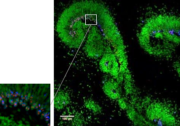
Neural Stem Cells in human developing brain
Dividing neural stem cells (green) in human fetal brain tissue (gestational week 18). Mitotic spindle is showed in red. This picture was performed with a wide spinning disk using a 40X water immersion objective: it is a 3D mosaic of 3×3 fields of view, 37 stacks with a step size of one micron. Then the mosaic was reconstructed in 3D and a maximum projection was finally processed.