Spinning disk
Frap/Photoablation

 Over the last decade, fast confocal spinning disk system has overcome classical raster confocal system for live imaging since it combines high speed with low photobleaching. Equipped with state of the art scientific camera spinning disk microscopy is able to achieve up to 1000 fps. It can also be combined with various devices to extend its capabilities for doing: Live super resolution, photoablation, FRAP/PA, ring TIRF.

Competition for Space Induced Cell Elimination through Compaction-Driven ERK Downregulation

Moreno, E., Valon, L., Levillayer, F. & Levayer, R.
Competition for Space Induced Cell Elimination through Compaction-Driven ERK Downregulation. Curr Biol

APPLICATIONS

In depth live tissue wounding

Drosophilia pupal Notum wounding performed with a Gataca systems’ spinning-disk equipped with an iLas-pulse for photo-ablation. In green : Z-projection of E-cad::GFP. In purple : Z-projection of a live sensor of ERK. Photo-ablation was done using a UV pulsed laser on 400x400x10 px hollow square. The two channel stacks were acquired every 2min and combined with an autofocus between each time point. 

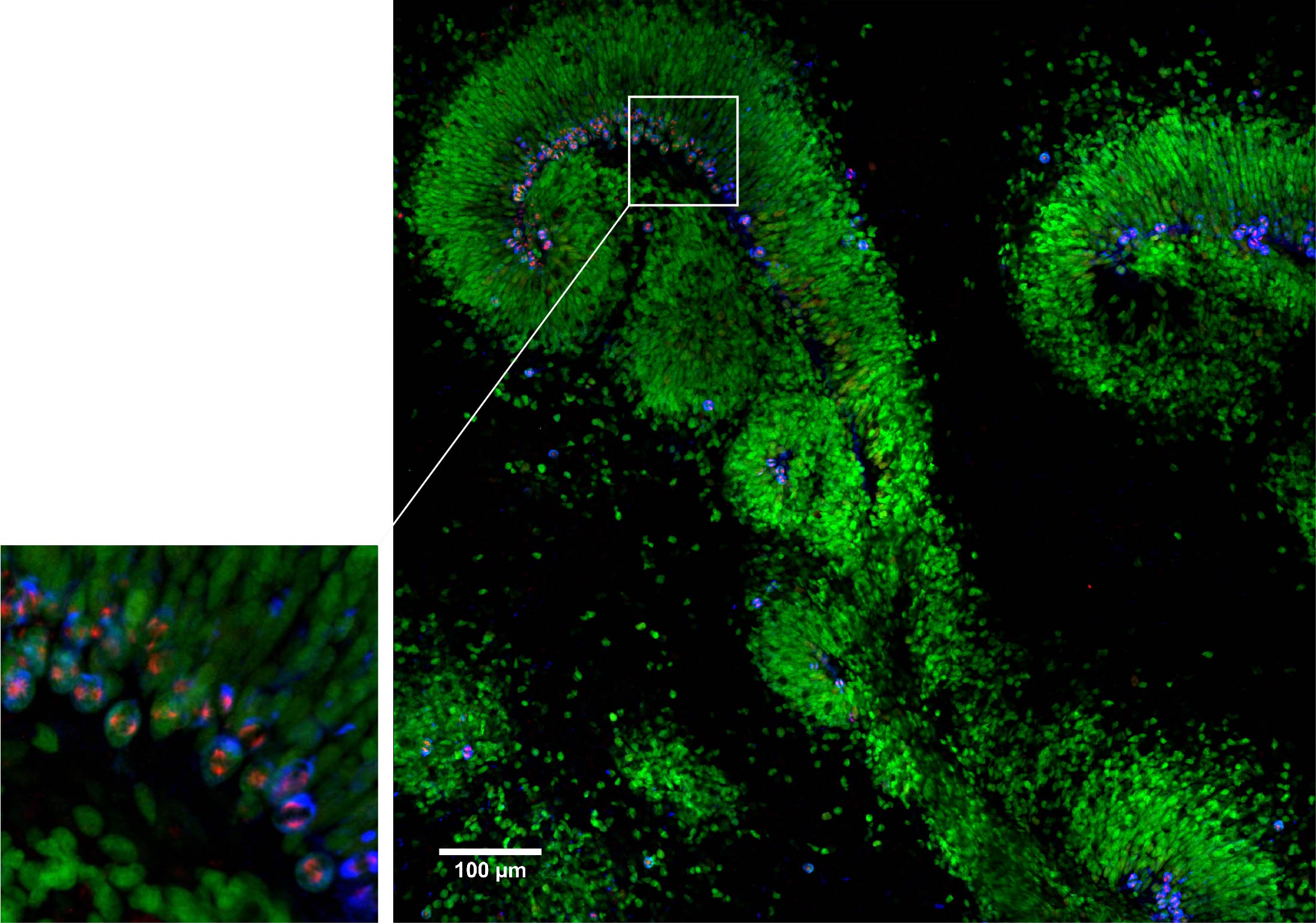
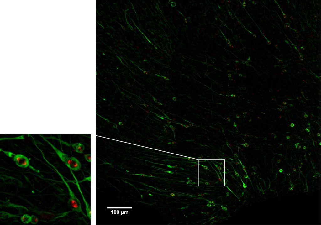
Neural Stem Cells in a human forebrain organoids

Cerebral organoids are derived from human induced pluripotent stem cells. Those mini brains in 3D show multiple lumens that recapitulate the organization of the developing cortex. Neural stem cells (green) form a neuroepithelium that surround the lumens. Stem cells divisions occur mostly at the lumen border, with condensed DNA (blue) and mitotic spindle (red). This picture was performed with a Gataca systems’ spinning disk using a 40X water immersion objective: it is a 3D mosaic of 3×3 fields of view, 17 stacks with a step size of two microns. Then the mosaic was reconstructed in 3D and a maximum projection was finally processed.
Courtesy of L. Coquand, A. Baffet’s lab, UMR144 CNRS/Institut Curie, Paris.

Neural Stem Cells in human developing brain

Dividing neural stem cells (green) in human fetal brain tissue (gestational week 18). Mitotic spindle is showed in red. This picture was performed with a wide spinning disk using a 40X water immersion objective: it is a 3D mosaic of 3×3 fields of view, 37 stacks with a step size of one micron. Then the mosaic was reconstructed in 3D and a maximum projection was finally processed.
Courtesy of L. Coquand, A. Baffet’s lab, UMR144 CNRS/Institut Curie, Paris.

Membrane trafficking

Fast live imaging of eYFP-Languerin (pH sensitive) tranfected HeLa cells. Single basal plane 20fps. With Live-SR super resolution module in by-pass mode.

Courtesy of J. Salamero’s lab, UMR144 CNRS/Institut Curie, Paris.

Related Products

Yokogawa CSU-X1

Spinning Disk

Confocal Systems

iLas2

iLas 2

Gataca Products

iLas3-400

iLas 3

Gataca Products

Gataca iLas Pulse

iLas Pulse

Gataca Products